1914 Nobel Prize in Physics
Reason for Award
for his discovery of the diffraction of X-rays by crystals
Laureates
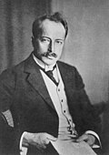
 German Empire
Explanation
X-rays are very energetic light that doctors use to see our bones. Max von Laue shone these X-rays onto a tiny salt crystal. The rays spread out into many directions just like ripples on water hit a rock. This spreading pattern is called “diffraction.” Laue’s experiment showed that X-rays behave like waves and that atoms inside a crystal are arranged in a regular order. Because of this, people found a new way to study the invisible arrangement of atoms.
Related Keywords
X-ray
X-rays are electromagnetic waves with much shorter wavelengths and higher energy than visible light. Discovered by Wilhelm Röntgen in 1895, they have been widely used for medical imaging and materials analysis. Their 0.01–10 nm wavelength range is comparable to interatomic distances, making them ideal for crystal structure studies. X-rays penetrate most substances but are strongly absorbed by heavy elements such as lead. Modern synchrotron and free-electron-laser sources provide extremely bright X-rays, enabling time-resolved measurements and nanoscale imaging. Laue’s work demonstrated the wave behaviour of X-rays, vastly expanding their scientific applications.
crystal diffraction
Crystal diffraction is the interference scattering that occurs when X-rays, neutrons or electrons interact with the ordered atomic arrays of a solid. Constructive interference arises when the phase difference of incident and scattered waves matches a lattice vector, producing discrete spots or rings. From spot positions we derive lattice constants, and from intensities we infer atomic coordinates and thermal motion. X-ray crystallography is indispensable for determining protein and drug structures and has led to many Nobel Prizes in Chemistry. Diffraction data are converted via Fourier transforms into electron-density maps, which are refined into atomic models. Laue’s discovery marked the historical starting point of this method and has supported materials science for over a century.
crystal lattice
A crystal lattice is a mathematical array of points representing the periodic arrangement of atoms or molecules in three dimensions. Each point is equivalent, and the entire structure can be generated by translating one point with lattice vectors. Specifying the unit cell’s shape and contents fully describes the crystal’s symmetry and structure. Laue diffraction spots map reciprocal-lattice vectors and therefore give direct information on real-space lattice constants. In solid-state physics, this periodic structure determines electronic band structures and phonon dispersions. Defects and dopants locally disturb the lattice, creating functionalities exploited in advanced materials and semiconductor devices.
Bragg's law
Bragg’s law, expressed as 2d sinθ = nλ, links the interplanar spacing d, incident angle θ, and wavelength λ for constructive interference. It was derived in 1913 by W. H. and W. L. Bragg to account for the angular positions of X-ray diffraction peaks. Whereas Laue used polychromatic radiation, Bragg’s law provided the theoretical basis for monochromatic angle-scan measurements. The simple formula applies universally, from large unit cells of proteins to nanocrystals. It works both in fixed-wavelength variable-θ experiments and in fixed-θ variable-λ setups. Modern powder X-ray and neutron diffractometers still employ Bragg’s condition as the first step in data analysis.
Laue spots
Laue spots are small bright dots recorded on a photographic plate when a crystal is illuminated with polychromatic X-rays. Each spot corresponds to a point in reciprocal space, and together they form symmetry patterns reflecting the crystal’s point group. By analysing the spot arrangement one can instantly determine crystal orientation, lattice system and symmetry operations. The method is still used for orientation analysis of metals and minerals and for quality assessment during single-crystal growth. With ultrafast pulsed X-rays, nanosecond stress changes can be tracked as shifts in Laue spots, enabling time-resolved measurements. Laue spots provide the most direct evidence of the wave interaction between X-rays and crystals demonstrated by Laue.
Ewald sphere
The Ewald sphere is a geometric construction that visualizes diffraction conditions in three-dimensional reciprocal space, drawn as a sphere of radius 1/λ. Placing the tail of the incident wave vector at the sphere’s centre, diffraction occurs when a reciprocal-lattice point touches the sphere’s surface. The diagram gives an intuitive grasp of the possible θ angles and the multiple diffraction conditions under polychromatic illumination. The Ewald sphere is a universal tool employed in electron, neutron and even phonon scattering analyses. Computational simulations sweep the sphere’s interior to predict diffraction intensities rapidly in dynamic modelling. By spatially expressing the Laue condition, the Ewald sphere complements Laue’s theory and forms a foundational concept in crystallography education.