1906 Nobel Prize in Physiology or Medicine
Reason for Award
for their work on the structure of the nervous system
Laureates
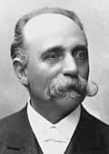
 Kingdom of Italy
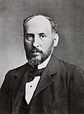
 Spain
Spain
Explanation
Our body has a "cable network" in which the brain and nerves exchange signals that travel like electricity. More than 100 years ago, Mr. Golgi and Mr. Cajal used a special chemical to turn nerve cells black and saw their shapes clearly for the first time. Thanks to that, they could see the long thread-like axons and the tree-branch-shaped dendrites extending from each cell. They showed that each nerve cell is separate and passes messages at its tips. This discovery became the model for diagrams in today’s textbooks and helps doctors understand brain diseases.
Related Keywords
Golgi staining method
The Golgi staining method uses a two-step chemical bath to impregnate only a small subset of neurons with dark silver chromate. Unlike previous global stains, it allows individual dendrites and axons to be followed without overlap. Developed by Golgi in 1873, it is often called the "black reaction". Cajal optimized the protocol’s timing and temperature, achieving high-resolution images especially in juvenile tissue. The technique became the starting point for modern connectomics and 3-D reconstruction approaches.
Neuron doctrine
The neuron doctrine states that the nervous system is composed of discrete cellular units called neurons. Information flows unidirectionally from dendrites to axons, and neurons are apposed at synapses but not physically continuous. Supported by Cajal’s anatomy and Sherrington’s physiology, it formed the core of the 1906 Nobel recognition. Electron microscopy and patch-clamp recordings subsequently confirmed the concept, making it the foundation of modern neuroscience. Although modified by discoveries of neuron-glia interactions and electrical synapses, the doctrine remains the standard framework.
Reticular theory
The reticular theory, dominant in the 19th century, proposed that the nervous system is a continuous network with no cellular boundaries. Many neurologists, including Golgi, favored it because the continuous appearance of stained processes seemed to support it. Although useful for explaining fast electrical phenomena, it was rejected after Cajal’s meticulous observations. The advent of electron microscopy, which revealed synaptic clefts between cells, provided conclusive evidence against it. Today the theory is remembered mainly for its historical role, though partial continuity exists in electrical synapses.
Dendrite
Dendrites are tree-like extensions from the neuronal soma that receive vast numbers of synaptic inputs. Cajal drew tiny protrusions on them, now called spines, hinting at structural plasticity. Dendritic morphology varies greatly among neuron types and determines how inputs are integrated. Calcium imaging and electrophysiology show local spikes and back-propagating action potentials within dendrites. Changes in spine density are reported in developmental disorders and brain diseases, making dendrites a potential diagnostic biomarker.
Axon
An axon is the single long projection of a neuron that carries electrical signals rapidly to distant targets. Cajal identified the axon initial segment and demonstrated that the axon is the output side of neuronal communication. In myelinated axons, saltatory conduction greatly increases propagation speed. Studies of Wallerian degeneration and inhibitory molecules after axonal injury inform therapies for nerve trauma. Axonal transport deficits are now implicated in diseases such as ALS and Alzheimer’s.
Synapse
A synapse is the junction where neurons exchange information, separated by a gap of only tens of nanometers. Named by Sherrington, it complements Cajal’s neuron doctrine. In chemical synapses, neurotransmitters are released and bind to receptors, producing voltage changes. Electrical synapses use gap junctions that allow ions to flow directly, enabling rapid synchrony. Synaptic plasticity, exemplified by long-term potentiation and depression, is considered the cellular basis of learning and memory.
Golgi apparatus
The Golgi apparatus is a membrane organelle that modifies and sorts proteins and lipids inside cells. It was discovered by Golgi in 1898 using silver impregnation. Composed of stacked cis and trans cisternae, it serves as a relay station for vesicle trafficking. Studies of COPI/COPII-coated vesicles clarified its function and led to the 2013 Nobel Prize awarded to Rothman and colleagues. In neurons, dispersed Golgi outposts in dendrites are thought to participate in local protein synthesis.
Neurohistology
Neurohistology is the discipline that studies the structure of the nervous system using histological techniques. It expanded dramatically at the end of the 19th century thanks to Golgi and Cajal’s achievements. Starting with light-microscope staining, it now encompasses electron microscopy, fluorescence imaging, and tissue clearing methods. Applications range from neuron classification and circuit mapping to clinical pathology. As new visualization technologies emerge, neurohistological knowledge continues to be renewed.