1967 Nobel Prize in Physiology or Medicine
Reason for Award
for their discoveries concerning the primary physiological and chemical visual processes in the eye
Laureates
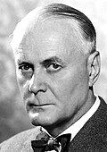
 Sweden
 United States of America
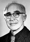
United States of America
Explanation
When we see something, light enters the eye and hits the “retina” at the back. The retina is packed with tiny sensors called “rods” and “cones” that change light into electrical signals and send them to the brain. Ragnar Granit, Haldan Keffer Hartline and George Wald studied exactly how this light-to-electric change happens. They found which sensors detect color and which ones help us see in the dark. Their work gives clues for curing eye diseases and for making better digital cameras.
Related Keywords
retina
A neural tissue lining the inside of the eyeball that converts light into electrical signals. It is stratified into layers containing photoreceptors, bipolar cells, ganglion cells and others. The retina performs the first stage of visual information processing, enhancing contrast and detecting motion. The laureates’ work measured individual retinal responses and clarified these functional divisions. Understanding the retina is essential for ophthalmic therapy and artificial-retina engineering.
rod cell
Approximately 100 million rods per eye specialize in scotopic (low-light) vision. They respond to near-single-photon stimuli and define the scotopic sensitivity curve. Rods contain rhodopsin as their principal pigment and have higher sensitivity than cones around 555 nm. The laureates analyzed rod electrical responses, explaining the mechanisms of dark adaptation. Their data underpin night-vision testing and the design of low-light imaging devices.
cone cell
Cones mediate photopic vision and color perception; humans have roughly six million. L, M and S types are sensitive to long-, middle- and short-wavelength light, respectively. The laureates measured cone spectra, establishing the physical basis of trichromatic color vision. Cone dysfunction causes color vision defects and acuity loss. Their findings influence display technology and clinical color-vision tests.
rhodopsin
A purplish visual pigment in rods composed of opsin and retinal, classified as a GPCR. Light converts its chromophore from 11-cis to all-trans, activating transducin and the phototransduction cascade. Wald clarified its spectral properties and vitamin-A-dependent regeneration. Rhodopsin defects cause congenital night blindness and retinitis pigmentosa. It is a target for drug development and optogenetic light-switch devices.
visual pigment regeneration
The biochemical cycle that restores bleached visual pigment via vitamin A metabolism. In the retinal pigment epithelium, all-trans retinol is isomerized back to 11-cis and recombines with opsin. Wald’s experiments proved this pathway, explaining night blindness caused by vitamin A deficiency. Delays in regeneration impair dark adaptation. The knowledge directly informs nutrition science and retinal therapy strategies.
lateral inhibition
A process in which neighboring neurons suppress each other’s activity, enhancing sensory contrast. Hartline quantified it in the horseshoe crab eye, providing a foundation for the center-surround receptive-field model of the retina. Lateral inhibition sharpens edge representation and increases information-compression efficiency in vision. Its dysfunction can cause hypersensitivity or reduced contrast perception. The principle is emulated in image-processing algorithms.
photoreceptor
A cell that converts light stimuli into neural activity. In animals these include rods and cones in vertebrates and R-cells in insect ommatidia, among others. The laureates pioneered microelectrode recordings from single photoreceptors, revealing their sensitivity and temporal properties. Photoreceptor dysfunction is a major cause of retinal degenerations. Artificial photoreceptor development is a focus in vision-restoration medicine.
electrophysiology
A discipline that measures and analyses electrical activity in living systems. Techniques such as microelectrodes and patch clamp record membrane potentials to study ion channels and synaptic dynamics. Granit and Hartline used early suction-electrode methods to record receptor potentials, establishing new analytical standards. Electrophysiology is widely applied in neuroscience, cardiology and muscle physiology. Advanced signal processing and machine learning are increasingly used for data analysis.
spectral sensitivity
The distribution of sensitivity to light across wavelengths. Cones show a tri-peaked curve, while rods have a single peak. The laureates matched absorption spectra to electrical responses, establishing cell-based theories of color vision. Measuring spectral sensitivity is vital in display design and lighting engineering. In vision research, genetically modified animals are used to analyze sensitivity shifts.
phototransduction
The molecular cascade converting light into electrical signals. Classic steps are rhodopsin activation → transducin → phosphodiesterase → cGMP decrease → closure of CNG channels → photoreceptor hyperpolarization. Wald’s pigment studies identified the initial chemical step of this cascade. Defects cause visual impairment and photosensitive seizures. Phototransduction principles are applied in gene therapy for light sensitivity and in biosensor development.