1924 Nobel Prize in Physiology or Medicine
Reason for Award
for his discovery of the mechanism of the electrocardiogram
Laureates
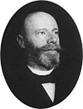
 Netherlands
Explanation
Our hearts beat thousands of times every day. Each beat sends a tiny electric signal through the body. Dr. Einthoven discovered how to draw this electricity as a line on paper, inventing the electrocardiogram (ECG). An ECG is like a picture of the heart’s activity that doctors can get just by placing wires on your arms and legs. Thanks to this, doctors can check the heart’s rhythm without opening the chest. Today ECG tests are even done at school check-ups and have saved many lives.
Related Keywords
electrocardiogram (ECG)
An electrocardiogram (ECG) records the heart’s electrical activity through electrodes placed on the skin. From the shapes of the P wave, QRS complex, and T wave clinicians evaluate sinus rhythm, ventricular hypertrophy, and arrhythmias. Because it is non-invasive and quick, it is widely used in emergency transport and routine check-ups. Digital analysis now enables real-time ST monitoring and prolonged Holter recordings. The fundamental principles devised by Einthoven more than a century ago still underpin clinical practice today.
string galvanometer
The string galvanometer stretches a thin, metal-coated quartz filament in a magnetic field and uses the Lorentz force on the current to deflect the string. Its high sensitivity allows optical amplification and recording of sub-millivolt potentials. Compared with prior support galvanometers, it had lower inertia and superior high-frequency response. This instrument enabled the first quantitative measurement of human cardiac potentials. Modern piezoelectric and fiber-optic sensors trace their design philosophy directly back to this historic device.
Einthoven's triangle
Einthoven’s triangle is an imaginary equilateral triangle formed by the two arms and the left leg, modelling the body-surface distribution of cardiac potentials. The three sides correspond to standard limb leads I, II, and III, which project the cardiac vector onto a plane. The geometric model yields the relationship I + III = II, allowing internal consistency checks of measurements. The triangle evolved into the basis for vectorcardiography and mean electrical axis analysis. Though simple, it remains essential in ECG interpretation and underlies the calculation of central terminals in modern 12-lead systems.
cardiac conduction
Cardiac conduction is the propagation of action potentials from the sinoatrial node through the atria, atrioventricular node, His bundle, and into the ventricles. The speed and pathways of conduction determine heart rate and stroke volume. Conduction abnormalities cause blocks and tachyarrhythmias, producing characteristic ECG changes. Einthoven’s instrument captured these signals with high temporal resolution, advancing understanding of conduction mechanisms. The concept remains pivotal in electrophysiological studies and pacemaker therapy today.
arrhythmia
Arrhythmia is a broad term for abnormal heart rhythms, including premature beats and atrial fibrillation. Before the ECG, diagnosis relied only on palpation and subjective symptoms, making detailed classification difficult. The ECG allowed electrical detection of the site, frequency, and duration of abnormal beats. This made it possible to recognise lethal ventricular fibrillation early and perform defibrillation, greatly improving survival. Today, AI-assisted long-term ECG analysis enables early detection of asymptomatic arrhythmias.
lead system
A lead system refers to the arrangement of electrode pairs used to record an ECG. Combining standard limb, precordial, and augmented leads allows three-dimensional visualization of the heart. Proper lead placement is essential for localizing myocardial infarction and confirming pacemaker electrode positions. Einthoven’s triangle is considered the foundational concept in lead system design. Recently, simplified 1–3-lead configurations for wearable devices have been developed, supporting telemedicine.
depolarization and repolarization
Depolarization is the change of membrane potential from negative to positive, forming the rising phase of an action potential. In cardiomyocytes it is triggered by the influx of sodium and calcium ions, leading to contraction. Repolarization is the return of the membrane potential to its resting value via potassium efflux, allowing relaxation and readiness for the next beat. On the ECG, the QRS complex represents ventricular depolarization and the T wave reflects repolarization. When these processes are delayed by ion-channel defects or drugs, the risk of serious arrhythmias such as long QT syndrome increases.