1938 Nobel Prize in Physiology or Medicine
Reason for Award
for the discovery of the role played by the sinus and aortic mechanisms in the regulation of respiration
Laureates
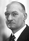
 Belgium
Explanation
When we breathe, tiny “guards” inside our body watch how much oxygen we have. Dr. Heymans discovered that these guards are in the big neck artery (the carotid artery) and in the chest’s big artery (the aorta). When oxygen is low, they tell the brain, and the brain makes us breathe faster or deeper. In short, he uncovered an important secret about how our body knows when to breathe.
Related Keywords
carotid sinus
A slight bulge at the carotid bifurcation that houses both pressure-sensing baroreceptors and chemoreceptors detecting blood gases. Sensory information travels mainly via the glossopharyngeal nerve to the nucleus tractus solitarius in the medulla. The processed signal alters vagal and sympathetic outflow, adjusting respiratory rate and heart rate within seconds. Clinical carotid-sinus massage exploits this reflex to terminate certain tachyarrhythmias, illustrating the translational impact of basic research.
aortic body
Small nodular structures near the aortic arch with potent chemosensory capabilities. Highly sensitive to hypoxia and pH shifts, their activation travels via vagal afferents to the respiratory centers, driving increased ventilation. Persistence of chemoreflexes after carotid sinus denervation highlights their complementary role. They remain under investigation as critical players in the pathophysiology of COPD and neonatal apnea.
chemoreceptor
Sensory organs that detect chemical constituents in blood or tissue fluid. Located peripherally in carotid and aortic bodies and centrally on the ventrolateral medulla. They rapidly sense hypoxia, hypercapnia, and acidosis, acting as major drivers of ventilatory command. Essential for increased breathing at high altitude and for maintaining respiratory rhythm during sleep.
baroreceptor
Mechanoreceptors that monitor arterial wall stretch and thus real-time blood pressure. Located mainly in the carotid sinus and aortic arch, their afferent signals alter heart rate and vascular resistance via the cardiac vagal reflex. Crucial for understanding hypertension pathogenesis and for maintaining blood pressure upon postural changes; research has informed the design of baroreflex activation devices.
respiratory center
A neuronal network in the medulla and pons that generates inspiratory and expiratory rhythms. Integrates input from chemoreceptors and higher brain areas, sending commands to the diaphragm and intercostal muscles. Suppression by drugs or disease can cause apnea, so monitoring its function is essential in intensive care.
glossopharyngeal nerve
Cranial nerve IX. Involved in taste and swallowing, and carries afferent fibers from the carotid sinus and body. Thus, it conveys chemo- and baroreceptor signals to the medulla to regulate breathing and circulation. Nerve injury can lead to impaired baroreflexes and dysphagia.
vagus nerve
Cranial nerve X. Provides parasympathetic control, slowing heart rate and enhancing gut motility, and transmits afferent information from aortic bodies and aortic arch. Vagal nerve stimulation is used to treat refractory epilepsy and depression, highlighting its broad clinical value.