1979 Nobel Prize in Physiology or Medicine
Reason for Award
for the development of computer assisted tomography (CT)
Laureates
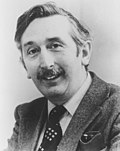
 United Kingdom of Great Britain and Northern Ireland
 United States of America
Explanation
When doctors need to look inside the body, they often use a special camera called a CT scanner. The CT sends X-rays through the body from many angles and a computer puts the information together to create slice-like pictures. In 1979, Mr. Hounsfield and Mr. Cormack built this system and received the Nobel Prize. Thanks to their work, doctors can see the brain or the belly without cutting the patient open and can find injuries or diseases quickly. This invention is one reason we can get fast and safe help when we are hurt.
Related Keywords
computed tomography
Computed tomography (CT) creates cross-sectional images by rotating X-rays around the body and mathematically reconstructing the data. The images are produced through an inverse Radon transform and depict not only bones but also soft tissues and vessels. Compared with MRI or ultrasound, CT offers high spatial resolution, and with contrast agents it can visualize blood flow dynamics. In emergency care, CT is indispensable for the rapid diagnosis of brain hemorrhage or multiple trauma. Ongoing improvements in dose management and reconstruction algorithms continue to push the technology forward.
X-rays
X-rays are high-energy electromagnetic waves discovered by Röntgen in 1895 and are capable of penetrating matter. The fraction of photons absorbed or scattered depends on tissue density and atomic number. In CT, measuring this attenuation enables image reconstruction, so the X-ray energy spectrum and filtration directly affect image quality. Dose is evaluated in milligray, requiring well-designed protocols and radiation protection measures. Recently, spectral detectors have exploited energy dependence for quantitative analysis.
image reconstruction algorithm
An image reconstruction algorithm is the mathematical procedure that converts projection data into pixel-based density values. Early CT relied on filtered back projection, which balanced speed and image quality. Today, iterative reconstruction using statistical modeling reduces noise and patient dose simultaneously. Hybrid techniques incorporating deep learning are emerging to suppress artifacts and achieve super-resolution. Choosing the proper algorithm affects diagnostic accuracy, acquisition cost, and computation time.
detector array
A detector array is a set of sensors that convert X-ray photons into electrical signals and is central to CT image quality. Energy-integrating designs using scintillators and photodiodes have been standard, but photon-counting arrays are gaining traction. Quantum efficiency and aperture size directly affect noise and resolution, making advances in materials science critical. Multislice CT stacks multiple rows of detectors along the z-axis, enabling the capture of several slices with each rotation. Future digital detectors with high temporal resolution are expected to refine the analysis of cardiac and arterial dynamics.
filtered back projection
Filtered back projection (FBP) is an analytic reconstruction method that filters the projection data in frequency space before back-projecting it. In the 1970s, when computing power was limited, FBP enabled near real-time image generation. The ramp filter preserves resolution by enhancing high-frequency components but also amplifies noise, making filter selection crucial. Extensions to fan-beam and cone-beam geometries allow FBP to remain the backbone of multislice and CBCT systems. Although iterative and deep learning reconstructions outperform it in noise characteristics today, its mathematical clarity still underpins physical understanding and scanner design.
Hounsfield Unit
The Hounsfield Unit (HU) is a normalized scale for CT gray values that sets water to 0 and air to −1000. HU is calculated as (µtissue − µwater) / µwater ×1000, allowing quantitative comparison of small density differences. Bone measures around +1000, fat around −100, and such ranges are used to diagnose abnormal shadows or to assess contrast enhancement. Differences in energy spectra or calibration among scanners can affect HU reproducibility. Spectral CT aims to correct energy dependence, enabling further standardization of HU and improved material differentiation.